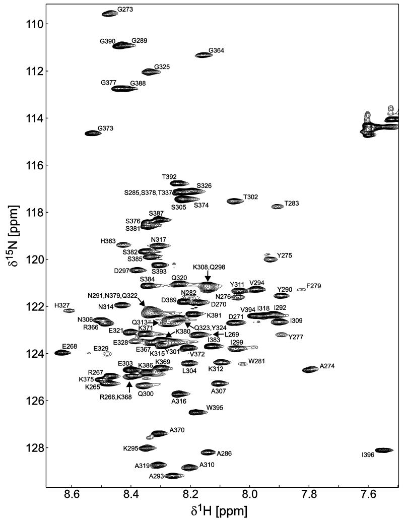
Figure 1. 15N-HSQC spectrum of the Cx45CT dimer in phosphate-buffered saline (pH 5.8) at 25°C.
Peak assignments for the backbone amides are indicated with numbering corresponding to the full-length Cx45 protein. The amide peaks that represent the dimerization domain (A333-N361) are not present in the spectrum due to the cross-peaks broadening beyond detection.

