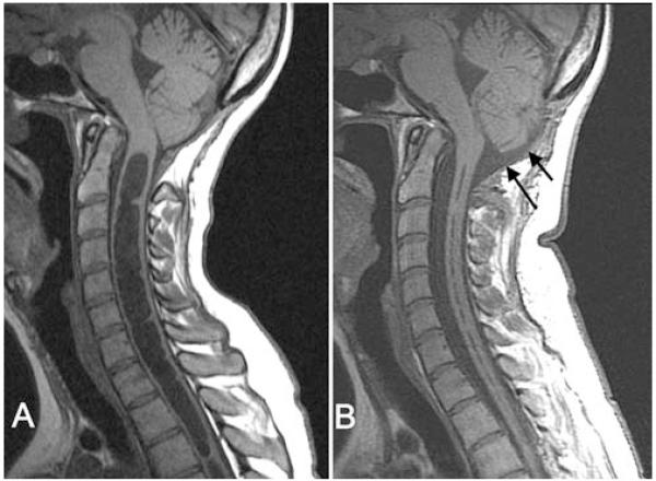
Fig. 1.
Midsagittal T1-weighted MR images demonstrating the posterior fossa and cervical spine before (A) and after (B) reexploration craniocervical decompression. After reexploration (B), the subarachnoid space at the foramen magnum expanded (arrows), the tips of the cerebellar tonsils rounded, and the syrinx diameter decreased dramatically.

