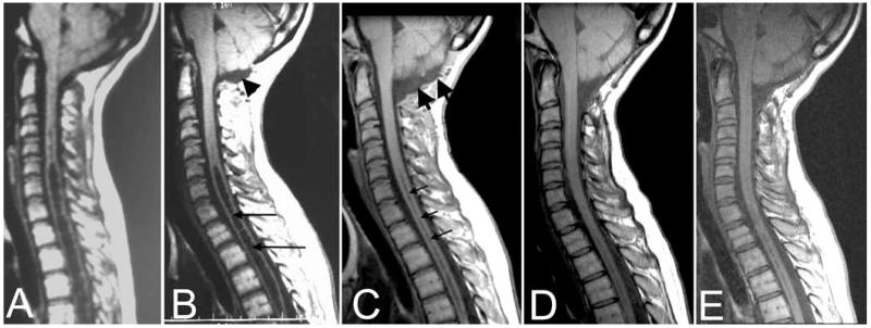
Fig. 6.
Midsagittal T1-weighted MR images of the posterior fossa and cervical spine before (A) and after (B) initial craniocervical decompression and then 3 months (C), 5 years (D), and 10 years (E) after reexploration surgery. After the initial surgery (B), myelopathy progressed and the syrinx (long arrows) did not resolve despite the creation of a CSF space dorsal and inferior to the cerebellar tonsils (arrowhead). At reexploration surgery, a pseudomeningocele was found dorsal to the dura. Reexploration craniocervical decompression removed the pseudomeningocele, enlarged the subarachnoid space at the foramen magnum (C, thick arrows), and led to syrinx resolution (C, thin arrows) that appears to be permanent (D and E).

