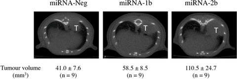
Fig 2.

Impact of TFPI-2 silencing on tumour progression in a nude mice orthotopic model. The intrabronchial implantation of cells was performed with a 1.9 Fr × 50 cm blunt-ended catheter that was inserted and advanced into the right main bronchus. Position of the catheter was monitored using X-ray imaging. Tumour cell suspension (7.5 × 10⁵ miRNA-Neg, miRNA-1b and -2b tumour cells in 25 μl) containing a 99mTc-labelled tin colloid and 10 mM EDTA was slowly injected into the right lobe. The scintigraphic assessment of the cell deposition into lung was then performed. Tumour progression was monitored using computed tomography scanning imaging over a 4-week period. Twenty minutes prior imaging, 100 mg/kg D-luciferin was injected i.p. to anesthetized mice. Lung tumour (T) volumes were measured on axial transverse sections. Each scanning image is representative of results obtained in nine animals per group. Tumour volume results are mean ± S.E.M. (n= 9).
