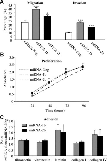
Fig 3.

Lung cancer cell migration, invasion, proliferation and adhesion to ECM proteins. (A) Cell migration and invasion were evaluated by modified Boyden chamber assay using culture inserts (8 μm pore size) coated with a thin layer of Matrigel (0.8 mg/ml) for invasion and uncoated inserts for migration. Cells (2 × 10⁵) were seeded in the upper chamber of the inserts and allowed to migrate for 48 hrs to the lower chamber containing culture medium with 10% FCS used as chemoattractant. Results from six experiments (means ± S.E.M.) are expressed as the percentage of migrating miRNA-1b and -2b cells compared with miRNA-Neg cells (*P < 0.05; **P < 0.001; ***P < 0.0001, Student t-test). (B) The proliferation potential of 1.25 × 10⁴ lung cancer cells transfected with miRNA targeting TFPI-2 was evaluated at 24, 48, 72 and 96 hrs using MTS assay. Results are means (±S.E.M.) of four experiments performed in triplicate for each time-point (P-values were determined using a Student t-test). (C) Tumour cells (2 × 10⁵) transfected with miRNA targeting TFPI-2 were allowed to adhere to fibronectin, vitronectin, laminin, collagen I and collagen IV or BSA used as control for 2 hrs and then stained with crystal violet. Absorbance readings measured at 570 nm with BSA were subtracted from ECM protein readings. Results from eight experiments performed in triplicate are expressed as the ratio of absorbances measured with miRNA-1b and -2b cells on values obtained with miRNA-Neg cells (means ± S.E.M.).
