
Fig 2.
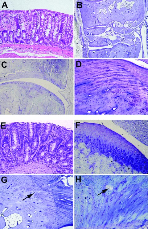
Microscopic features of disease in anti-TNF-α-treated HLA-B27 transgenic rats (B27TR). (A)–(D) Early anti-TNF-α treatment. (A) Histological section of colon in early-treated B27TR (haematoxylin and eosin staining). (B), (C) Representative histological sections of peripheral joint, with well preserved articular components. (D) Normal organization of the enthesis, with regular tidemarks and aligned chondrocyte rows towards the ligament. (B–D: toluidine blue staining). Original magnification: (A, C) ×10; (B) ×4; (D) ×40. (E)–(H) Late anti-TNF-α treatment. (E) Histological section of colon in late-treated B27TR (haematoxylin and eosin staining). (F)–(H) The histopathological joint features of three out of six late anti-TNF-α treated B27TR are shown. The other three late treated-B27TR showed no significant joint alterations as in the early treated group (B–D). (F) Areas of superficial matrix pallor, with hypertrophic chondrocytes and undulating articular surface. (G) Irregular tidemark between fibrocartilage and bone and endochondral bone ossification (arrow). (H) Bone erosion at the enthesis (arrow). [(F–H) toluidine blue staining]. Original magnification: (E–G) ×20; (H) ×40.