
Fig 4.
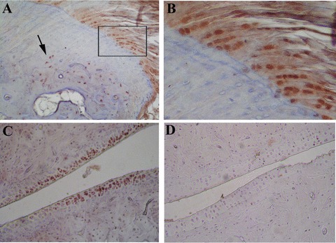
Representative immunostaining for TNF-α in the peripheral joints of HLA-B27 transgenic rats (B27TR). (A), (B) 18-week-old IgG2a,k-treated B27TR. (B) is a higher magnification view of boxed area in (A). Immunopositive chondrocytes in the fibrocartilaginous point of entheseal attachment (B) and in the proliferating zone at the border between bone and cartilage (arrow). (C) 27-week-old IgG2a,k-treated B27TR: immunopositive proliferating chondrocytes in the articular cartilage. (D) 18-week-old anti-TNF-α treated B27TR: absence of TNF-α immunopositivity in the cartilage. Original magnification: (A), (C), (D) × 20; (B) × 60.