
Fig 5.
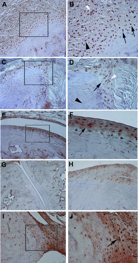
Representative immunohistochemical results of phosphorylated Smad1/5/8 in the peripheral joints of HLA-B27 transgenic rats (B27TR). (A), (B) 18-week-old IgG2a,k treated B27TR. (B) is a higher magnification view of boxed area in (A). Smad1/5/8 signalling is detected in spindle-shaped entheseal fibroblast-like cells (black arrowhead), inflammatory cells (white arrowhead), and in round chondroblast-like cells (black arrows) at entheseal level. (C), (D) 27-week-old IgG2a,k treated B27TR. (D) is a higher magnification view of boxed area in (C). Proliferating (white arrowhead) and prehypertrophic (black arrow) chondrocyte-like cells show phosphorylated Smad1/5/8-immunopositivity in the fibrocartilage at the point of entheseal attachment. Hypertrophic chondrocytes are negative (black arrowhead). (E), (F) 27-week-old IgG2a,k treated B27TR. (F) is a higher magnification view of boxed area in (E). Immunopositive proliferating chondrocytes are evident in the articular cartilage (black arrow). (G), (H) 18-week-old anti-TNF-α treated B27TR. Absence of phosphorylated Smad1/5/8 immunopositivity in the articular cartilage (G) and at the entheseal level (H). (I), (J) 27-week-old anti-TNF-α treated B27TR. (J) is a higher magnification view of boxed area in (I). Proliferating chondrocytes in the fibrocartilaginous point of entheseal attachment show strong immunopositivity for phosphorylated Smad1/5/8 (black arrow). Original magnification: (G) ×10; (A, C, E, H, I) ×20; (B, D, F, J): ×40.