
Fig 5.
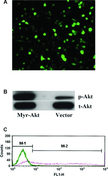
Transfection of MAPCs with Akt plasmids. After 24 hrs of transfection, the majority of MAPCs (over 60%) were successfully transfected with the constitutively active Akt plasmids as determined by fluorescence microscopy and cytometry as well as Western blot. (A) A fluorescence image of the transfected cells (20×) showing the majority of the cells are positive for the plasmids. (B) Phosphorylated Akt level was dramatically increased in the cells after transfection with Akt vectors as determined by Western blot. (C) Akt transfection efficiency in MAPCs was evaluated by flow-cytometry. The blue line presented the cells in the control group (in the M-1 Zone) that showed no positive expression of eGFP; whereas the red line demonstrated that over 60% of the cells (in the M-2 zone) were positive for eGFP after Akt transfection.