
Fig 6.
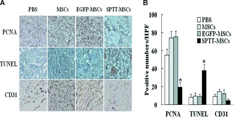
Immunohistochemical analysis of proteins related to cell proliferation, apoptosis, and the formation of neovasculature. For immunohistochemical analysis of tumour cell proliferation, apoptosis and microvessel density, tumours were paraffin embedded and sectioned. (A) Staining was performed with anti-PCNA antibody to assess proliferation, while TUNEL staining was performed to examine apoptosis, and a CD31 antibody was employed to detect endothelial cells, representing sites of vascularization. Slides were lightly counterstained with haematoxylin. (B) Quantitative analysis of proliferation (PCNA) and apoptotic (TUNEL) indices were calculated by counting positive cells in 10 random fields at 40× magnification. The number of proliferating tumour cells decreased, whereas the number of apoptotic cells increased in the group treated with SPTT-MSCs. Blood vessel counts were determined by counting the number of vessels in 10 randomly chosen areas of CD31 stained sections (40×). The number of CD31+ cells was decreased in the group treated with SPTT-MSCs (*P< 0.05 compared to mock-transduced MSCs).