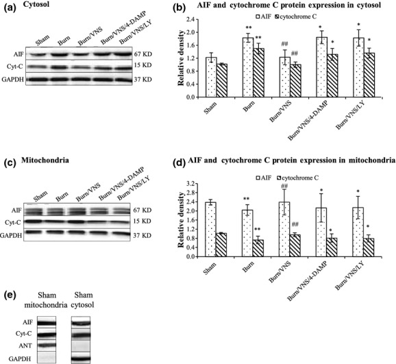
Fig. 5.

Western Blot Analysis of AIF and Cytochrome C Expression in Mitochondrial and Cytosolic Fractions of Heart Tissue. Cytosolic (A) and mitochondrial (C) fractions of heart tissue were isolated at 24 hrs after burn and underwent Western blot analysis for AIF and cytochrome C expression. ANT and GAPDH blotting were used as loading controls for mitochondrial and cytosolic proteins respectively. The density of the AIF and cytochrome C blot was normalized against that of GAPDH (B) and ANT (D) to obtain relative blot density. ANT was not detected in cytosolic fractions and GAPDH was not detected in mitochondrial fractions respectively (E). *P < 0.05 versus Sham, **P < 0.01 versus Sham; #P < 0.05 versus Burn, ##P < 0.01 versus Burn.
