
Fig 1.
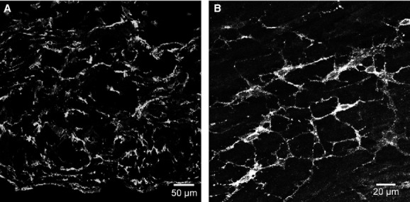
Platelet-derived growth factor receptor (PDGFR)-α cells in the bladder detrusor. (A) Cryostat section of detrusor muscle showing PDGFR-α immunoreactivity throughout the entire mucularis. (B) Whole mount of the bladder muscularis. PDGFR-α+ cells run parallel in the direction of the smooth muscle cells in the detrusor. PDGFR-α+ cells possess spindle- and stellate-shaped morphology with multiple processes that formed a discrete network. Scale bars are indicated in each panel.