
Fig 1.
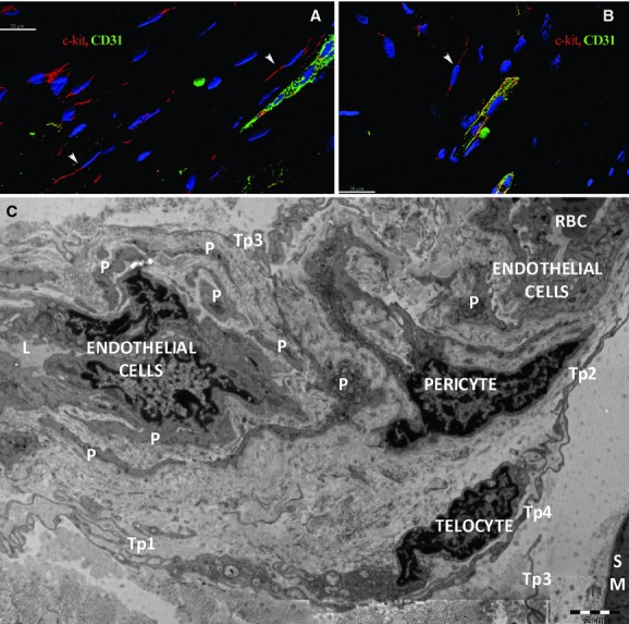
Human skeletal muscle; laser scanning confocal microscopy; three-dimensional shadow projection image. Double immunofluorescence labelling shows CD117/c-kit-positive cells (red) distributed around small blood vessels (arrowheads) (A) or capillaries (B) visualized by CD31 endothelial marker (green) in the perimysial and endomysial interstitial spaces. Nuclei are counterstained with 4′,6-diamidino-2-phenylindole (blue). Original magnification: 600×. (C) Electron microscopy. A telocyte with two telopodes (Tp1, Tp2) is located next to a pericyte which is visible in a twist of a capillary. Small fragments from pericytes (P) border the capillary. The basal lamina edges the pericytes. Telopodes (Tp3, Tp4, Tp5) belong to other telocytes.