
Fig 5.
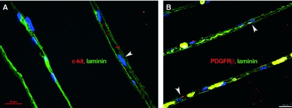
Human skeletal muscle; laser scanning confocal microscopy; basal laminae are visualized by laminin expression (green). c-kit-positive cells (A) and pericapillary platelet-derived growth factor receptor-β-positive cells (B) are clearly located in the interstitial space (arrowheads), between two adjacent skeletal muscle fibres enclosed by basal laminae. Nuclei are counterstained with 4′,6-diamidino-2-phenylindole (blue).