
Fig 2.
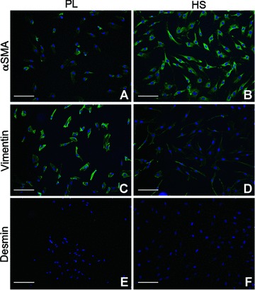
Immunofluorescent staining of MF cultured in PL and HS. Results for phenotyping of one patient are displayed, but results were comparable for all patients. (A, B) α-SMA, (C, D) vimentin and (E, F) desmin. Nuclei are stained with Hoechst. Original magnification was 10×. Bars represent 200 μm.