
Fig 4.
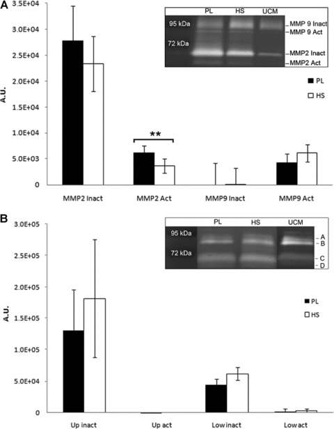
(A) Quantification of MMP2 and MMP9 expression by cells in culture (mean ± S.D.), corrected for unconditioned medium (UCM). Cells cultured in both media produce and activate MMP2, whereas MMP9 did not show a difference compared to the unconditioned control. Activated MMP2 expression was significantly lower in the cells cultured with HS (**P < 0.01). The inlay shows an example of expression patterns by one patient and the size of the signals that were quantified. A.U.: arbitrary units as a measure for number of counted pixels in a pre-determined surface. (B) Quantification of collagenases by cells in culture (mean ± S.D.). Expression patterns are presented in the inlay. We did not find significant differences in collagenase expression between cells cultured in PL or HS. Up: MMPs expressed at marks A and B; Low: MMPs expressed at marks C and D. Active forms (Act, B and D), are slightly smaller in size when compared to inactive forms (Inact, A and C) of these MMPs.