
Fig 2.
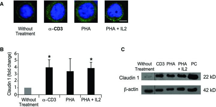
Claudin 1 is up-regulated in activated PBLs. PBLs from healthy volunteers were activated in vitro with either anti-CD3 antibody (α-CD3), PHA, PHA and IL2 or left without treatment (non-activated) and analysed by (A) immunofluorescence analysis with anti-claudin 1 antibodies (green) and a nuclear dye (TO-PRO 3 – blue). Scale bar: 5 μm. (B) Western blot analysis of claudin 1 protein levels. Bar graph depicts the fold change of claudin 1 protein levels relative to cells without treatment. The protein levels of claudin 1 were normalized to β-actin levels for each activation protocol using densitometry analysis. The data are presented as the mean ± S.E. of three independent experiments. *P = 0.04 (Student’s t-test). (C) Representative Western blot of claudin 1 expression in PBLs. PC: a positive control of extracts from HEK 293 cells transfected with a claudin 1 expression vector.