Fig. 6.

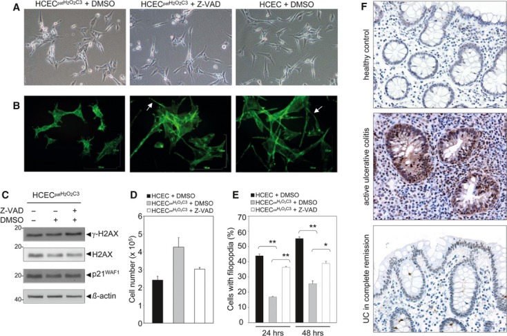
Restoration of the normal HCEC phenotype through caspase inhibition. (A) Phase contrast micrographs showed that treatment of HCECpatH2O2C3 with the caspase inhibitor Z-VAD-FMK led to the restoration of the morphological HCEC phenotype after 24 hrs. (B) FITC-Phalloidin-staining of HCECpatH2O2C3, HCECpatH2O2C3 treated with Z-VAD-FMK and HCEC. Filopodia are marked. (C) HCECpatH2O2C3 were treated with caspase inhibitor Z-VAD-FMK, and protein expression of γ-H2AX and p21WAF1 was analysed after 72 hrs. An increase in both γ-H2AX and p21WAF1 expression was detected. (D) Cell numbers of HCEC, HCECpatH2O2C3 and Z-VAD-FMK-treated HCECpatH2O2C3 after 72 hrs. The data are representative of three independent experiments. (E) Determination of percental filopodia-containing cells of HCEC and HCECpatH2O2C3 and of Z-VAD-FMK-treated HCECpatH2O2C3 after 24 and 48 hrs. Data indicate mean ± SEM and were obtained from two independent experiments. *P < 0.05, **P < 0.01. (F) Immunohistochemical analysis of p-JNK in normal colonic mucosa, active ulcerative colitis (UC) and UC in complete remission.
