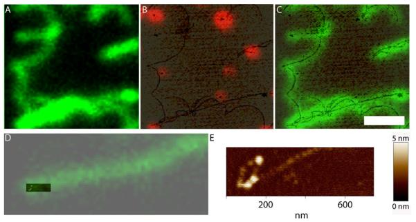
Figure 6.
Fluorescence human RAD51 protein filaments on DNA. A) Optical image showing green fluorescence filamentous structures originated by the association of the Alexa 488 conjugated RAD51 with the long DNA molecules of lambda phage (~48 kbp). Height image overlaid with the optical image from red fluorospheres (B) or green RAD51 (C) from the same region shown in A. D) Overlay of height and green fluorescence images of one filament end. E) Extra resolution in the height image shows a lariat at the end of this filamentous structure. Scale bar 2 μm.

