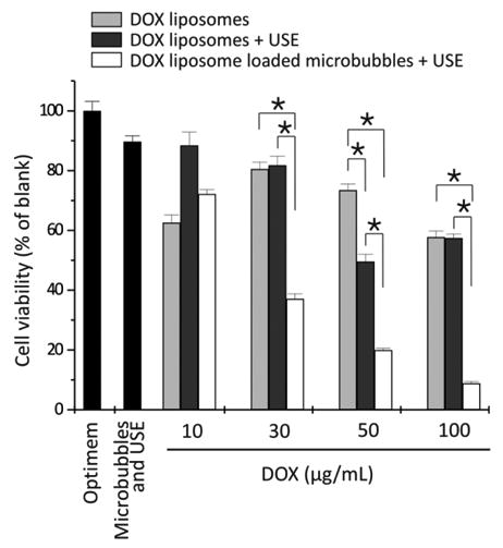
Figure 5.

Cell viability of the melanoma cells after treatment with DOX-liposomes without (light gray bars) and with ultrasound exposure (USE) (dark gray bars) and DOX-liposome-loaded microbubbles after ultrasound exposure (white bars) as a function of the DOX concentration in the OptiCell. For higher doxorubicin concentrations, more doxorubicin liposomes were used, whereas the microbubble concentration was always 109 microbubbles per OptiCell unit. *P < 0.05.DOX, doxorubicin. Reprinted by permission from Macmillan Publishers Ltd: Molecular Therapy, [113], copyright (2009).
