
Fig 3.
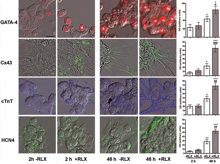
Representative merged differential interference contrast and confocal immunofluorescent micrographs of cardiomyocytes cultured for 2 and 48 hrs in the absence or presence of RLX. Immunoreactivities for the different antigens are shown in pseudo-colours. There is a visible increase in the immunoreactivity for the noted antigens upon a 48-hrs incubation with RLX. GATA-4–positive nuclei are indicated by asterisks. Bar (upper left) = 16 mm. The right panels show the computer-aided densitometry of the immunostaining for the noted myocardial marker proteins. Significance of differences (one-way ANOVA, n = 5): *P ‘ 0.05 and **P ‘ 0.01 versus −RLX 2 hrs; +P ‘ 0.05 and +++P ‘ 0.001 versus +RLX 2 hrs; #P ‘ 0.05, ##P ‘ 0.01 and ###P ‘ 0.01 versus −RLX 48 hrs.