
Fig 2.
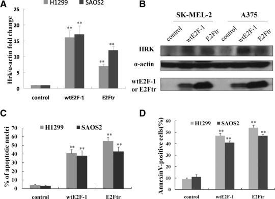
Up-regulation of HRK in wtE2F-1- and E2Ftr-induced apoptosis is independent of p53 status. H1299 and SAOS2 cells, which lack p53, were infected at MOI 100. After 24 hrs of infection, RT-PCR (A) and Western blot analysis (B) were performed as described in Figure 1B and C. After 48 hrs of infection, the percentage of apoptotic cells was counted by Hoechst 33258 staining under a fluorescence microscope (C) and determined by annexin V-PE staining by flow cytometry analysis (D). Values represent mean ± S.D. of three independent experiments (bars: mean ± S.D.; **P < 0.01 compared with control; n = 3).