
Fig 4.
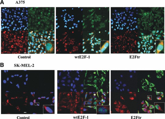
wtE2F-1 and E2Ftr are not directly acting to induce apoptosis at mitochondria. After 24 hrs of infection, A375 cells (A) and SK-MEL-2 cells (B) were stained with mitotracker red 580 and HRK (A) or E2F-1 (B) and then counterstained with DAPI (blue) (upper left panel) as described in Materials and methods (magnification, ×600). (A) An overlay (lower right panel) of the red (mitochondria, lower left panel) and the green (HRK, upper right panel) is provided in yellow to show co-localization of HRK in mitochondria. (B) wtE2F-1 or E2Ftr is primarily distributed in the nuclei and cytoplasma as shown in green (right upper panel). An overlay (lower right panel) of the red (mitochondria, lower left panel) and the green (wtE2F-1 or E2Ftr, right upper panel) is not shown in yellow, which suggests wtE2F-1 or E2Ftr is not located in mitochondria. Insets represent higher magnifications of the boxed areas (bar = 20μm).