
Fig 8.
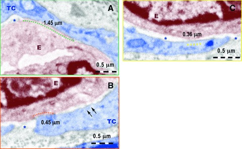
Higher magnification of the fields marked by colour rectangles in Figure 7. (A) A nanocontact of about 1.45 μm long between the telocyte (TC) and the membrane of the endothelial cell (E) is marked by the green dotted line. The asterisk indicates the intercellular space between TC and E. No endothelial basal lamina is present. (B) A nanocontact of about 0.45 mm long between the E and TC is indicated by the red dotted line. The asterisk mark intercellular space without endothelial basal membrane and the two arrows show possible electron dense ‘feet’ connecting the endothelial cell membrane and the TC membrane. (C) A nanocontact of 0.36 mm long between endothelial cell membrane and TC is indicated by yellow dotted line between the endothelial cell membrane and TC membrane. No basal lamina is visible in the extracellular space between the endothelium and TC (asterisks).