
Fig 5.
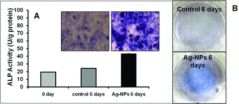
Effect of AgNPs on the ALP activity of MC3T3-E1 cells. (A) Microscopic images showing the enhanced level of ALP after 6 days after AgNP exposure. The cells were stained by ALP double staining and the level of the ALP enzyme was evaluated. (B) Actual images of the ALP stained Petri dishes for the control and the cells exposed to AgNPs and cultured for 6 days.