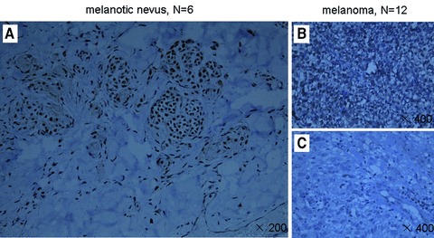
Fig 5.

Menin expression is reduced in certain primary melanoma cells. Sections from paraffin-embedded samples were stained with affinity-purified anti-menin antibody for immunohistochemistry staining. (A) Menin was easily detectable in the nucleus of the pigmented nerves (×200). (B and C) In melanoma, staining for menin was slightly weaker (three cases) or undetectable (nine cases), as compared to that in the pigmented nevus cells (×200).
