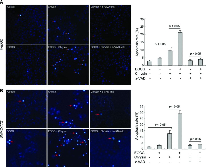
Fig 8.

EGCG potentiates chrysin-induced apoptosis. (A) HepG2 cells were treated with or without 20 μM EGCG, 10 μM chrysin and 20 μM z-VAD-fmk for 48 hrs. Apoptosis was assessed by Hoechst 33342 staining. Red arrow: representatives of apoptotic cells. Quantification of apoptotic cells was performed by taking the images in random fields and counting cells with strong fluorescence, condensed or fragmented nuclei. The apoptosis rate was plotted. Columns: mean percentage of apoptotic cells; bars: S.E. (B) SMMC-7721 cells were treated with or without 20 μM EGCG, 20 μM chrysin and 20 μM z-VAD-fmk for 48 hrs. Apoptosis was assessed by Hoechst 33342 staining. Red arrow: representatives of apoptotic cells. The apoptosis rate was plotted. Columns: mean percentage of apoptotic cells; bars: S.E. A representative of three experiments was shown.
