
Fig 8.
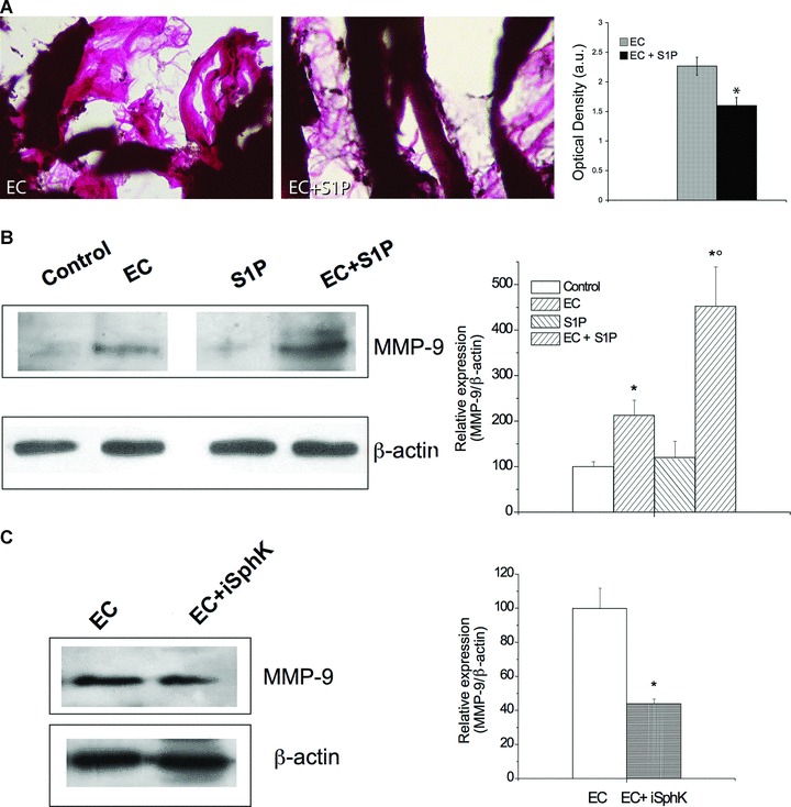
Effect of S1P on extracellular matrix remodeling and MMP-9 expression. (A) Light microscopy. Van Gieson stained muscle sections revealing collagen fibres (pink). In the histogram densitometric analysis of collagen staining intensity (Student’s t-test; *P < 0.05, n= 3). (B) Western blot analysis of MMP-9 protein expression in control, S1P-treated, EC- and EC + S1P-damaged muscle fibres. (C) MMP-9 protein expression in EC-treated cells in the absence and presence of SphK1 inhibitor (iSphK). Protein aliquots (30 μg) of tissue lysates were immunoblotted and detected by ECL. Band intensity was determined by densitometry and relative percentage to control arbitrarily normalized to 100 is shown in the graphs. (Student’s t-test; *P < 0.05 versus specific control, n= 3; °P < 0.05 versus EC).