
Fig 2.
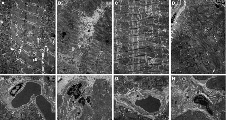
Histopathological examination of biopsies of left ventricular tissues. Representative electron micrographs of cardiomyocytes (upper panels) and blood capillaries (lower panels) from: sham-operated rats (A, E), showing normal mitochondria and myofibrils and microvessels with open lumina and normal organelle complement; rats undergoing 30 min. ischaemia followed by 60 min. reperfusion (B, F), showing mitochondrial swelling (asterisk), severe myofibril hypercontraction and constriction of microvascular lumen (arrow); rats subjected to I/R and treated with 1 mg/kg K5-N,OSepi (C, G), showing cytoplasmic oedema, moderate myofibril hypercontraction and nearly normal microvessels; rats subjected to I/R and treated with 1 mg/kg B4/100 (D, H), showing similar myocyte and endothelial alteration as in the untreated I/R rats. Bars = 1 μm.