
Fig 8.
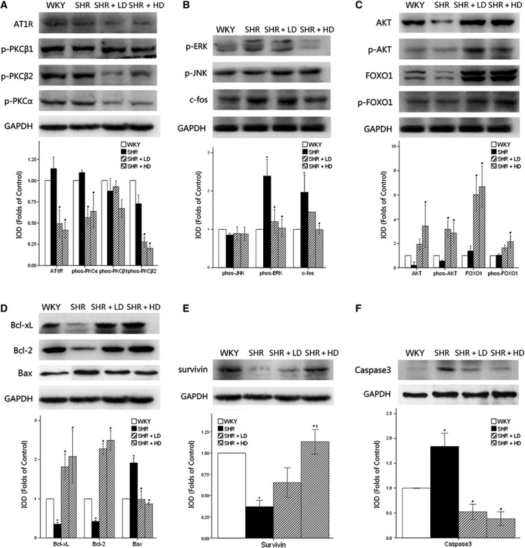
Western blot analysis of hypertrophy-associated signaling pathways and anti-apoptotic signaling pathways in myocardium of WKY controls, SHR controls, SHR + LD and SHR + HD; the relative level of integrated optical density (ΔIOD) measured. (A) the expressions of AT1 receptor, phos-PKCα and phos-PKCβ2 significantly down-regulated in SHR + LD and SHR + HD (P < 0.05); no significant changes in expression of phos-PKCβ1 (P > 0.05); (B) the expressions of phos-ERK and c-fos significantly up-regulated in SHR controls compared with WKY controls (P < 0.05), the former reversed in SHR + LD and SHR + LD, the latter reversed in SHR + LD (P < 0.05); the expression of phos-JNK remained consistent (P > 0.05); (C) Akt, phos-Akt, FOXO1 and phos-FOXO1 up-regulated in SHR + LD (P < 0.05); (D) the expressions of Bcl-xl and Bcl-2 significantly suppressed, and the expression of Bax significantly augmented in SHR controls compared with WKY controls (P < 0.05), and reversed in SHR + LD and SHR + LD (P < 0.05); (E) survivin down-regulated significantly in SHR controls and reversed in SHR + HD (P < 0.05), with significant difference between SHR + LD and SHR + HD (P < 0.05); (F) Caspase-3 expression augmented in SHR (P < 0.05) and suppressed in SHR + LD and SHR + HD (P < 0.05). Values, mean ± SED; n = 3; *P < 0.05 versus WKY controls; #P < 0.05 versus SHR controls; δ P < 0.05 versus SHR + LD.