
Figure 1.
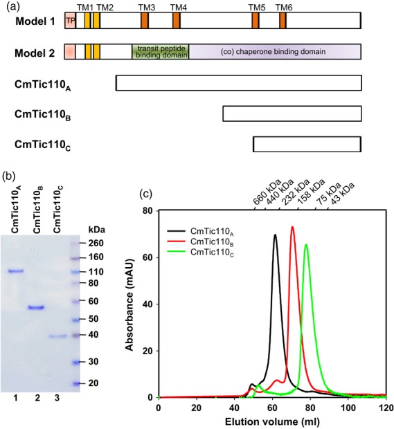
Purification of the three CmTic110 proteins. (a) Schematic representations of the two structural models of Tic110 and the corresponding regions covered by CmTic110A, CmTic110B and CmTic110C. Locations of the proposed transmembrane domains (TM1 and TM2 are represented by yellow boxes; TM3 to TM6 are represented by orange boxes), transit peptides (TP, pink boxes) and domains for transit-peptide binding (green box) and (co) chaperone binding (purple box) are marked. (b) CmTic110 proteins were purified to homogeneity. One microgram of purified proteins was analyzed by SDS-PAGE and stained by Coomassie blue. (c) Purified CmTic110 proteins were analyzed by gel filtration. The chromatograph of each CmTic110 protein is shown.