
Figure 5.
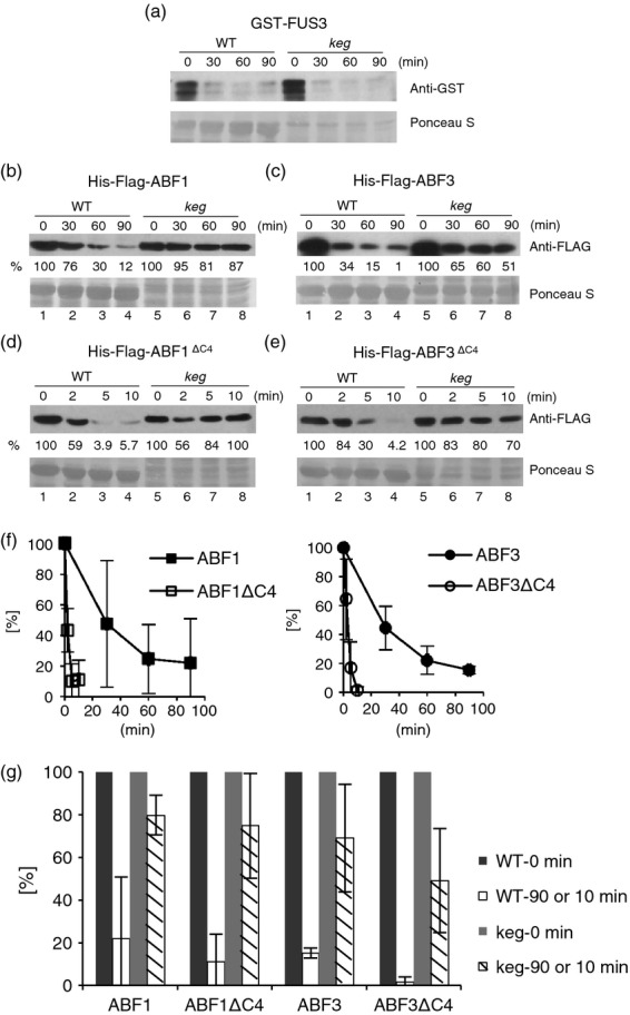
In vitro degradation of ABF1 and ABF3 is slowed in keg.Bacterially expressed recombinant GST-FUS3 (a), His-Flag-ABF1 (b), His-Flag-ABF3 (c), His-Flag-ABF1ΔC4 (d) or His-Flag-ABF3ΔC4 (e) was incubated with a 7–day-old Col or a 14–day-old keg seedling extract over the indicated time courses [note the shorter times in (d) and (e) compared with (b) and (c)]. His-Flag-tagged protein levels were visualized by anti-Flag immunoblotting. Ponceau S staining was used as the loading control. (f) Data from independent experiments comparing the in vitro degradation of full-length proteins and their respective ΔC4 forms were plotted normalized to time 0 (ABF1, n = 3; ABF3, n = 2).(g) Protein at 90 or 10 min for full-length and ΔC4 forms, respectively, were normalized to time 0. Student's t–tests indicate that the loss of the same protein was significantly slower in keg extracts compared with the wild type (WT; n = 3; P < 0.03, P < 0.02 and P < 0.03 for ABF1, ABF1ΔC4 and ABF3 ΔC4, respectively). (Test not performed for ABF3, this specific time course was repeated twice, and a different time course, with the same results was performed once.) Note: cannot compare full-length and ΔC4 forms here because time points are different (see Figures 5f and 6 for these comparisons).