
Fig. 3.
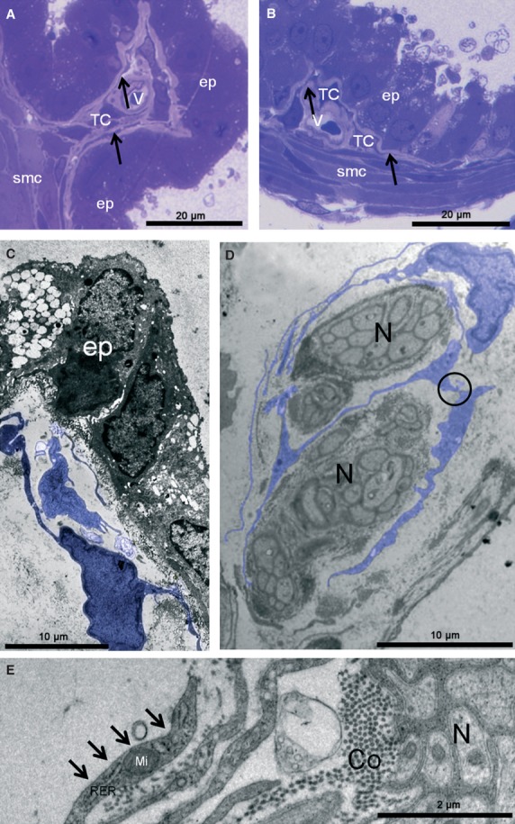
Prostate semi-thin sections for light microscopy and ultra-thin sections for transmission electron microscopy. (A and B) Detailed view of a telocyte (TC) and its elongations (arrows) in the subepithelial region. These images were obtained with light microscopy for 0.5-μm sections. (C–E) Ultrastructural features of prostate telocytes (TC). The distribution of this cell type was verified primarily in the subepithelial region. The TC is identified in these figures in blue (C and D). (D) Two telocytes close to prostatic nerves (N) showing extremely thin, long processes extending from the cellular body, with repeated curving. Black circle indicates a close contact between two TCs. (E) Detail of TC located in the perineural region. Note that the extensions of the TC are not observed basal lamina (arrows). ep (epithelium), smc (smooth muscle cell), v (blood vessel), RER (rough endoplasmic reticulum), Mi (mitochondria), TC (telocyte), N (nerve), Co (collagen).