5.

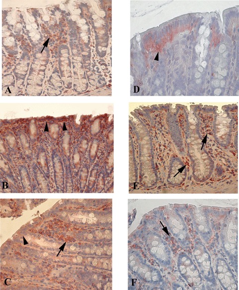
Immunohistochemistry for Fas and Fas-ligand (Fas-L) on colonic mucosal tissue. (A–C) Fas expression. (A) Fas-positive cells (arrow) in the lamina propria of non-transgenic F344 rats. (B) In IgG2a, k-treated B27TR, Fas-positive cells of the lamina propria significantly decreased, whereas positive intestinal epithelial cells (IEC) increased (arrowheads). (C) Anti-TNFα treatment increased significantly the number of Fas positive cells in the lamina propria (arrow), while the positive IEC decreased (arrowhead). (D, E) Fas-L expression. (D) In control non-transgenic F344 rats, Fas-L was expressed mainly by the IEC (arrowhead). (E) IgG2 a,k-treated B27TR showed a higher number of positive cells in the lamina propria (arrows). (F) Anti-TNFα treatment had no effect on Fas-L expression in the lamina propria (arrow) compared to IgG2a,k treatment. Red colour: AEC immunohistochemical developing; Blue colour: haematoxylin counterstain. Original magnification A, B, C, E× 20; D, F × 40.
