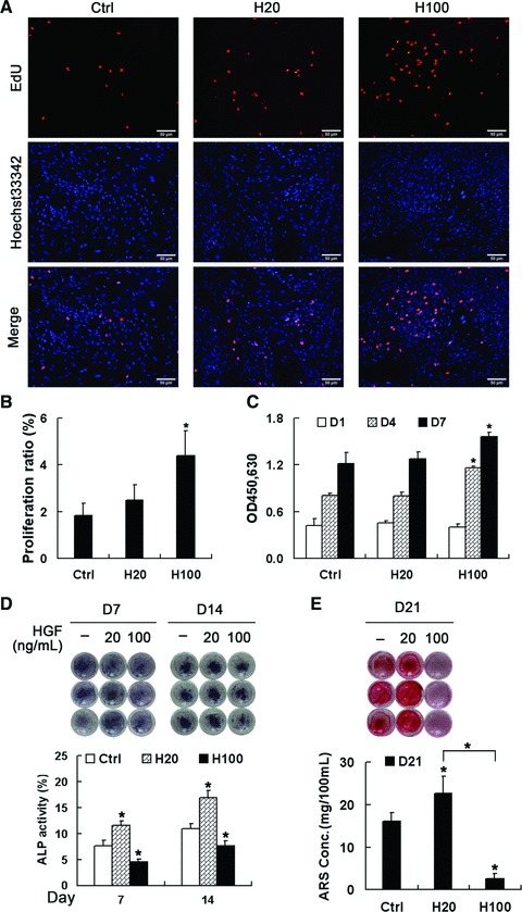
Fig 2.

Effects of HGF concentration on MSC proliferation and osteogenic differentiation. (A, B) EdU incorporation assays in MSCs after treatment with HGF for 24 hrs. Scale bar ∇ 50 μm. (C) Cell viability assays after treatment with HGF for 1, 4 and 7 days. Both assays indicate that H100 treatment more strongly induces proliferation compared with H20 treatment. (D, E) MSCs were maintained in DM containing HGF for 21 days and the level of osteogenesis was assessed at the indicated times by staining with NBT-BCIP to show ALP (D, upper panel) or with alizarin red sulfate (AR-S) to show calcium deposition (E, upper panel). (D and E, lower panels) Quantification analysis showed that H20 promotes differentiation, which was strongly suppressed by H100. *P < 0.05, compared with control group at each time point.
