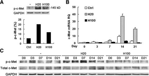
Fig 3.

Activation of c-Met was highly enhanced by H20, but suppressed by H100 during osteogenic induction. (A) Western blot analysis of MSCs left untreated (–) or treated with HGF using anti-phosphorylated-c-Met (top panel) or anti-GAPDH (as a loading control, bottom panel) antibodies. H20 treatment led to a much stronger activation of p-c-Met than H100 treatment beginning from 15 min. (B) Quantitative RT-PCR of c-Met mRNA expression during differentiation with long-term treatment of HGF. H20 treatment stimulated significantly higher levels of c-Met, which were partially suppressed by H100 treatment compared with control. (C) C-Met activation during differentiation by Western blot analysis is in consistency with short-term treatment with HGF. RQ: relative mRNA expression normalized to GAPDH.
