
Fig 7.
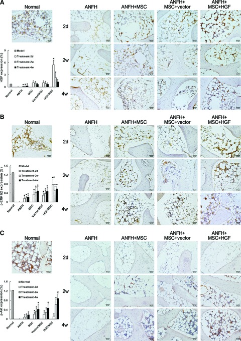
Immunohistochemical detection and semi-quantitative analysis of HGF expression (A), phosphorylation of ERK1/2 (p-ERK1/2) (B) and Akt (p-Akt) (C) in vivo after induction of hormone-induced ANFH and treatment with transplantation of MSCs. The level of HGF greatly decreased after induction of ANFH, but significantly increased 2 days after transplantation of MSCs, accompanied by increased p-ERK1/2. Two weeks post-transplantation HGF levels decreased, which was followed by a significant increase in p-Akt levels. The effects were most remarkable in the HGF-expressing MSC-treatment group. *P < 0.05, compared with the normal group. #P < 0.05, compared with the non-infected MSC-treated group. Scale bar ∇ 20 μm.