
Fig 1.
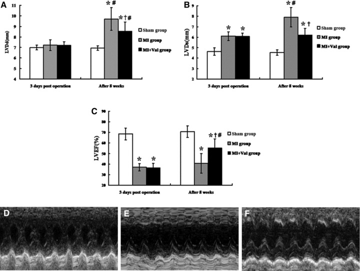
Echocardiography results and images of three groups. M-mode images were taken from the level of the LV outflow tract and the mitral valve was studied during the activity of ventricular wall, especial anterior wall. (A–C) The changes of LVDd, LVDs and LVEF 3 days after operation and after 8 weeks. The LV wall motion in Sham group (D) was better than the other groups. The condition of MI + Val group (F) was improved better compared with the MI group (E). LVDd: LV dimension end diastole; LVDs: LV dimension end systole; LVEF: LV ejection fraction; *P < 0.01 versus Sham group; †P < 0.01 versus MI group; #P < 0.05 versus 3 days after operation.