
Fig 1.
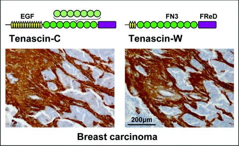
Tenascin structure and tumour stroma staining. A schematic model representing one subunit of the hexameric tenascin-C and tenascin-W proteins is shown above a breast carcinoma section stained with antibodies against the two proteins. Both tenascins are built from the following domains: a central, N-terminal oligomerization domain (black line), EGF-like repeats (EGF, yellow), FN type III repeats (FN3, green; FN3 repeats subject to alternative splicing in light green) and a C-terminal fibrinogen related domain (FReD, violet). Antibodies against tenascin-C and tenascin-W strongly stain the stroma of the breast carcinoma, although the epithelial tumour nests (nuclei in blue) are negative.