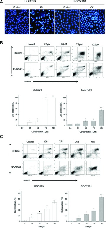
Fig 2.

CK induces apoptosis in human gastric carcinoma cells. (A) Effect of CK on the apoptotic morphology of human gastric carcinoma cells. Cells were stained with Hoechst-33342, and images were captured by fluorescence microscopy. Magnification, 100×. (B) Cells were treated with different concentrations of CK (0, 2.5, 5.0, 7.5 and 10.0 μmol/l) for 24 hrs. (C) Cells were treated with 5 μmol/l CK for different time periods (0, 12, 24, 36 and 48 hrs), and the apop-tosis was determined by annexin V/PI staining assay. Data are reported as means ± S.D. of three separate experiments. * and ** indicate P < 0.05 and P < 0.01 compared with control group, respectively.
