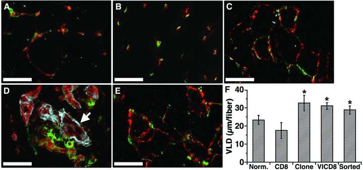
Fig 4.

FACS-purified rVICD8 myoblasts induce normal, stable and mature angiogenesis in ischemic muscle. (A–E) The medial hamstring muscles were harvested 3 months after myoblast injection and frozen sections were immunostained with antibodies against endothelium (PECAM, in red), pericytes (NG2, in green) and smooth muscle cells (α-SMA, in blue). Images were taken in areas where myoblast engraftment had been confirmed in adjacent serial sections stained with X-Gal. (A) nonischemic, non-treated muscle. (B) rICD8 control cells. (C) reference clone. (D) primary transduced rVICD8 population. (E) FACS-purified rVICD8 myoblasts. The white arrow in (D) indicates a large angioma-like aberrant structure. Size bars 50 7μm. (F) Vessel length density (VLD) was measured around engrafted fibres and is expressed as the mean vessel length (in m) per fibre. Values represent the mean ± S.E.M. (n = 3–5). CD8: control cells; VICD8: primary transduced rVICD8 myoblasts. *P < 0.05 compared to control cells.
