
Fig 3.
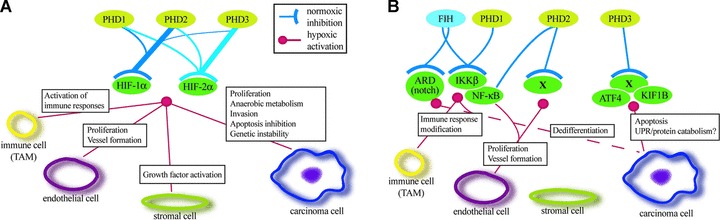
Schematic model of the HIF hydroxylase function that occurs in HIF-dependent (A) or HIF-independent manner (B). (A) The activities of different PHD isoforms towards the two HIF-α isoforms are illustrated by the line thickness. Main HIF-dependent functions of PHDs on different tumour cell types are indicated. (B) Some of the activities of the hydroxylases on potential non-HIF hydroxylation targets and their function on different tumour cell types. X indicates non-characterized hydroxylase targets.