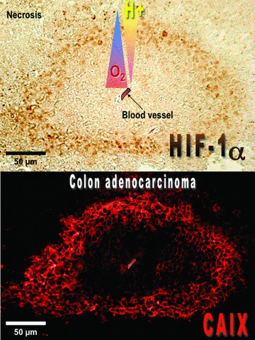
Fig 6.

Immunohistochemical analysis of HIF-1α and CAIX expression in mice xenograft tumours. Tumour sections obtained from subcutaneous injection of colon carcinoma LS174Tr cells were stained with an anti-CAIX monocalonal antibody M75 and showed correlation between CAIX expression and regions of HIF-1α+ staining. A relationship between CAIX localization and regions of hypoxia and necrosis, was also noted. Staining of CAIX is predominantly membranous while HIF-1α is localized in the nucleus. Magnification 20×.
