
Fig 1.
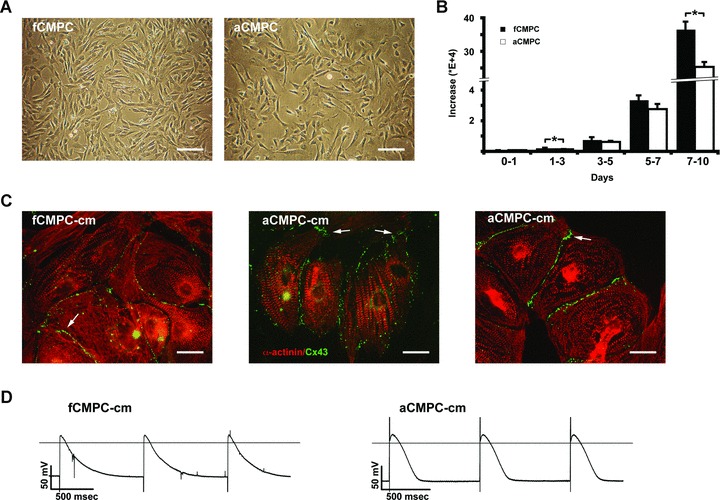
Proliferation and differentiation of CMPCs. (A) Bright field images showing foetal (fCMPC, left) and adult CMPCs (aCMPC, right) under normal culture conditions. Note the similarities in spindle-shaped phenotype. Scale bars: 200 μm. (B) Growth differences between fCMPCs and aCMPCs. Cells were counted at day 1, 3, 5, 7 and 10 after plating. A significantly higher increase in cell number was observed in fCMPCs versus aCMPCs between day 1 and 3 (P= 0.007, n= 3) and day 7 and 10 (P= 0.013). (C) Immunostaining for α-actinin (red) and Connexin 43 (green) in foetal (fCMPC-cm, left) and adult CMPC-derived cardiomyocytes (aCMPC-cm, middle and right). White arrows indicate the presence of gap junctions at the cell membrane borders. Scale bars: 50 μm. (D) Action potentials recorded from fCMPC-cm (left) and aCMPC-cm (right) using sharp microelectrodes. Monolayers were field stimulated at ∼1 Hz. Field stimulation artefacts (seen as a sharp downward peak at the onset of the fCMPC-cm action potentials and a sharp upward peak at the aCMPC-cm action potentials) did not affect the overall phenotype of CMPC action potential.