
Fig 3.
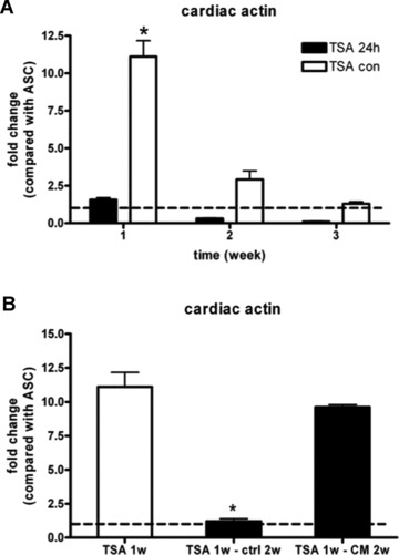
Real-time RT-PCR analysis of cardiac actin mRNA in hASCs after trichostatin A treatment for 24 hrs, and cultured in control medium or continuously for 1, 2 and 3 weeks (A). ASCs also treated by TSA continuously for 1 week were cultured in control medium or CCM for 2 weeks (B). GAPDH was used as internal control. Bar graphs indicate fold change of mRNA compared with ASCs (dotted line). Results are shown as mean ± S.E.M. (n= 3). *P < 0.05 versus other groups