
Fig 2.
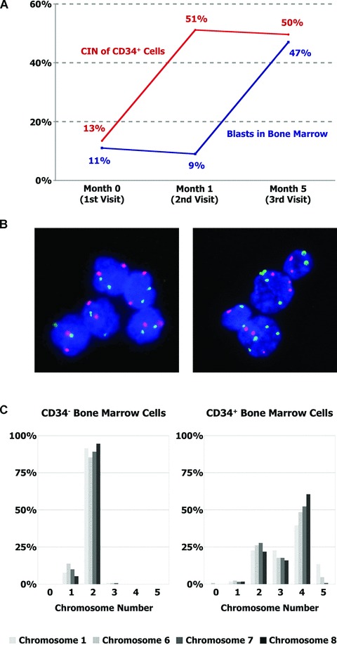
Increasing numerical CIN levels precede the progression to AML. (A) Bone marrow specimens from patient #15 (see Table 2), who initially presented with MDS, were obtained at the indicated time-points. Blasts were quantified by routine cytology, and numerical CIN levels in CD34+ haematopoietic progenitor cells were evaluated in the same specimens. A blast count exceeding 20% and thus progression to AML was first detected at month 5, while a massive rise in the CIN level was already apparent at month 1. (B) CD34+ cells of patient #15 at the first visit (left panel) and 1 month later (right panel), hybridized with centromeric FISH probes to chromosomes 6 (red) and 7 (green). DNA was counterstained with DAPI (blue). The increase in CIN preceded the evolution to secondary AML by 4 months. (C) The numerical CIN level was determined by evaluating the number of the indicated chromosomes per cell in at least 100 cells. Comparison of CD34-negative (left panel) and CD34-positive (right panel) bone marrow cells from patient #15 at month 5 revealed that numerical CIN was confined to CD34+ cells and equally affected all analysed chromosomes.