
Fig 3.
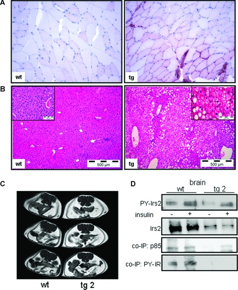
Histological analysis in muscle and liver tissues, MRT and insulin signalling in brain tissues in PKC-β2 transgenic and wild-type mice. (A) Muscle tissue was taken from wild-type (wt) and transgenic (tg) mice. Some fibres of the transgenic animal show intense lipid droplet accumulation using Oil Red O staining. Representative slides out of three animals are shown. (B) Histological analysis in liver tissues by haematoxylin and eosin stained liver sections in 6-month-old wt and tg mice. Hepatic steatosis was graded by lipid-loaded hepatocytes in three animals per genotype. (C) T1-weighted MR images of a 6-month-old wild-type (wt) or PKC-β2 transgenic (tg) mouse to detect intra-abdominal fat mass. (D) Western blot analysis of Irs2 immunoprecipitates detecting tyrosine phosphorylation (PY-Irs2) and expression in brain tissues of PKC-β2 and wild-type mice. Animals were stimulated intravenously for 5 min. with insulin or saline as a control. Tyrosine phosphorylation of Irs2 co-immunoprecipitated IR (PY-IR) and of p-85 was detected by using the respective antibodies. A representative immunoblot is shown out of three independent experiments, for quantification see result section.