
Fig 2.
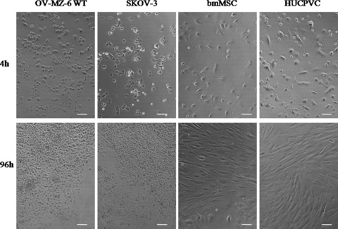
Light microscopical images of four cell types at different time-points. After 4 h of seeding 8300 cells/cm2 OV-MZ-6 and SKOV-3 cells are attached to the bottom of the cell culture plate; bmMSC are not fully attached yet as elongated fibroblast-like features are missing; HUCPVC show fibroblast-like morphology. After 96 h OV-MZ-6 and SKOV-3 cells proliferated and displayed a typical epithelial cobblestone formation; bmMSC showed slight proliferation and fibroblast-like features; HUCPVC showed typical morphological features (magnification 10×, scale bar 100 μm).