
Fig. 1.
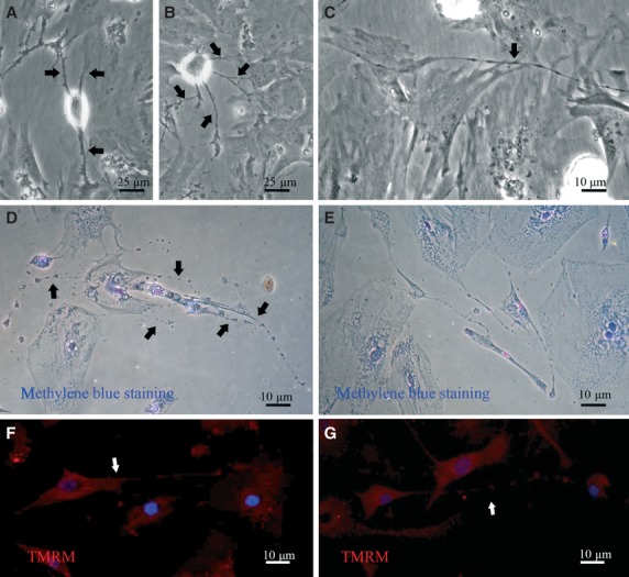
Telocytes in 2D breast stromal cells: phase contrast microscopy, supravital methylene blue staining and mitochondria-specific staining. (A) Fusiform TCs with long processes (indicated by arrow). (B) Polygonal TCs with four long, thin prolongations (indicated by arrows). (C) Triangular TCs with bead-like telopodes, eight dilations in telopodes were visible (indicated by arrows). (D) and (E) Supravital methylene blue staining of TCs. An octopus-like telocyte with five bead-like telopodes (indicated by arrows) in D. TCs connected with surrounding cells to form a network by their telopodes with bead-like conformation in E. (F) and (G) Mitochondria immunolabelled with TMRM of TCs. The strong prolongation with dilations of telocyte could be observed in primary breast stroma cell culture. The prolongations were indicated by arrows.