
Fig. 2.
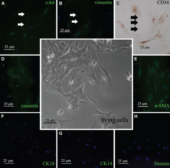
Immunofluorescence and immunohistochemistry of TCs and identification of mammary gland stromal cells in 2D culture. The central inset indicated stromal cells derived from breast tissue with different morphology of cells observed under phase contrast microscope. (A) Fluorescence microscopic observation showed c-kit/CD117-positive cells with obviously telopodes (green), and dilations were pointed by arrows. (B) Immunofluorescent staining result of vimentin-positive TCs with long prolongation (green, indicated by arrows). (C) Immunohistochemical staining result of CD34-positive TCs with telopodes. The dilations were indicated by arrows. (D–H) Immunofluorescent staining for other stromal cells in 2D monolayer cell culture. (D) vimentin-positive cells, (E) α-SMA-positive cells, (F) CK18-negative cells, (G) CK 14-negative cells, (H) Desmin-negative cells.